FIG. 6.
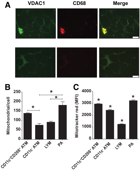
A: Crown ATMs and PAs are enriched for mitochondria. Formalin-fixed adipose section stained with antibodies against VDAC1 and CD68 or nonspecific antibodies. VDAC1 staining intensity was greatest for crown ATMs and less intense for resident ATMs and CD68-negative cells. Scale bar = 50 μm. B: Mitochondria per cell was calculated as the relative amount of mitochondrial versus genomic DNA, determined by qPCR of DNA isolated from sorted stromovascular cells using CYTB and LEP primers. Results are means ± SE of three independent experiments. *Significantly different (P < 0.05) in ANOVA by posttest comparison. C: Stromovascular cells were incubated in RPMI/10% FCS with or without 50 nmol/l Mitotracker red for 15 min in a 37° water bath and then incubated on ice with CD206FITC, CD11cPE, and CD45PC7 antibodies before analysis using flow cytometry. Results are means ± SE of three independent experiments. *Significantly different (P < 0.05) in ANOVA by posttest comparison. (A high-quality digital representation of this figure is available in the online issue.)

