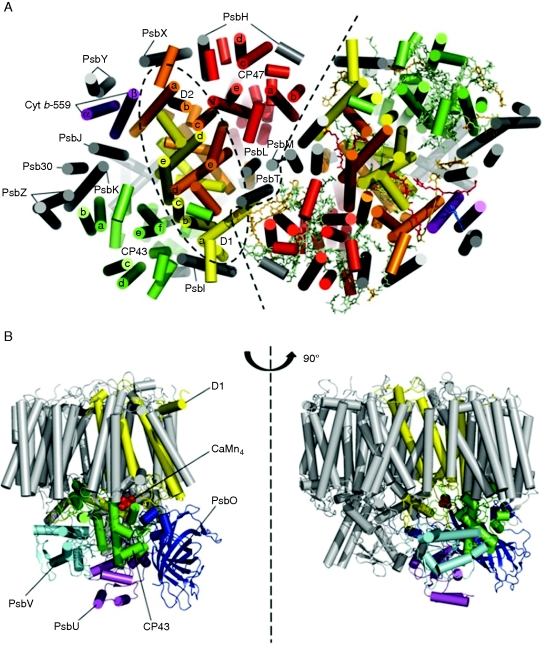
Fig. 1.
Sub-unit organization of the isolated homodimeric PSII complex from Thermosynechococcus elongatus. (A) View from the cytoplasmic side of the membrane. The two monomers are separated by a black dashed line and the α-helical elements of each subunit are represented as cylinders. D1 (yellow), D2 (orange), CP43 (green), CP47 (red), cytochrome b-559 (purple) and the remaining 11 small sub-units (grey) are indicated in the monomer on the left side as well as the D1–D2–Cyt b-559 sub-complex (elliptical black dashed circle). The same colour coding system applies to the monomer on the right side where are also represented the co-factors of PSII: chlorophylls (green), carotenoids (orange), pheophytins (yellow), plastoquinones (red) and haem (blue). The co-factors are shown in stick form. (B) Two side views, differing by a rotation of 90 °, showing the lumenal subunits PsbO (dark blue), PsbV (light blue), PsbU (purple) and the large lumenal loop of CP43 interconnecting transmembrane helices e and f (green) that lies close to the CaMn4 cluster. The figure was created with the software Pymol (http://pymol.sourceforge.net, version 0·99) and the PDB files 3BZ1 and 3BZ2 (Guskov et al., 2009).

