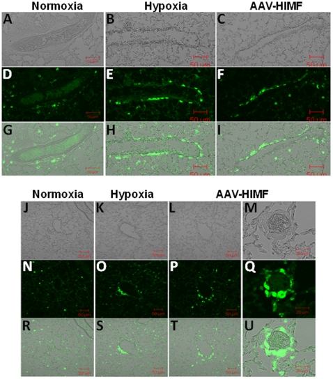
Figure 3. Both chronic hypoxia and pulmonary HIMF gene transfer recruit BMD cells to the pulmonary vasculature.
(A–C, J–M) Light micrograph of fluorescence images to show blood vessel structure. Frozen sections from normoxic (20.8% O2) (D, N), hypoxic (10.0% O2) (E, O), and AAV-HIMF treated (2.5×10¹⁰ VP) (F, P, Q) lungs were stained with a rabbit anti-GFP polyclonal antibody that was visualized by an FITC-conjugated goat anti-rabbit IgG antibody (green). (G–I, R–U): Differential interference contrast images of light and fluorescence images to show structure. A–L, N–P, R–T: Scale bar: 50µm. M, Q, U: Scale bar: 20µm.

