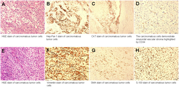
Figure 2.
Histopathologic pictures of the metastatic HCC. Hematoxylin and Eosin (H&E) staining showed tumor cells infiltrate in the bony trabeculae and soft tissue (A and E). There were two patterns of tumor cells (A and E). Histopathologic pictures of the metastatic HCC. Hematoxylin and Eosin (H&E) staining showed tumor cells infiltrate in the bony trabeculae and soft tissue (A and E). There were two patterns of tumor cells (A and E). The carcinomatous tumor cells with pleomorphic nuclei and abundant cytoplasm arranged in solid sheets and nests were positive for HepPar-1 and cytokeratin-7 (CK7) staining (B and C). The carcinomatous tumor cells also demonstrate sinusoidal vascular stroma highlighted by CD34 (D). The other sarcomatous cells with little cytoplasm arranged in the myxo-chondroid background were positive for vimentin, SMA, and S-100 protein staining (F to H). These features are compatible with a metastatic hepatocellular carcinoma with sarcomatous change. Magnification: 200×.

