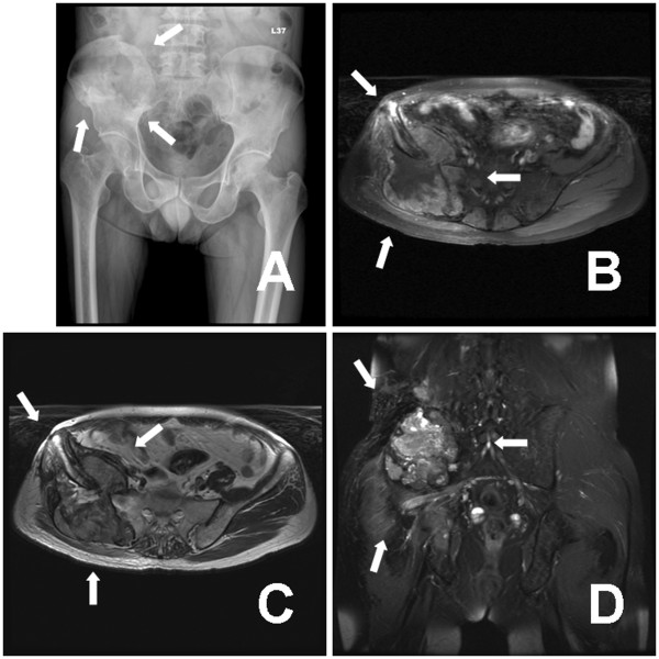
Figure 3.
Pelvis radiograph showed a large ill-defined osteolytic lesion at right iliac bone accompanied with iliac wing fracture (white arrows) (A). Repeated MRI revealed a 9.4 × 9.2 × 9.0 cm bipeduncular lesion arising from previous operation site, accompanied with iliac wing fracture (white arrows) (B to D). B: axial fast spin echo (FSE) fat-suppressed T1-weighted image with contrast enhancement. C: axial fast recover fast spin echo (frFSE) T2-weighted image. D: coronal fat-suppressed (FS) FSE T2-weighted image with contrast enhancement.

