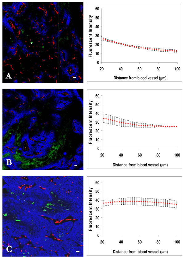
Figure 2.
Dose response distribution of cetuximab in relation to blood vessels and regions of hypoxia in A431 xenografts. Left panels show the distribution of cetuximab (blue) in relation to blood vessels (red) and regions of hypoxia (green) in A431 xenografts at 24 h after an i.p. injection of (A) 0.01 mg, (B) 0.05 mg, and (C) 1.0 mg. In right panels staining intensity (mean +/- SEM) due to cetuximab is plotted against distance from the nearest blood vessel in the tumor section. Note minimal drug binding in hypoxic regions. Scale bar = 100 μm.

