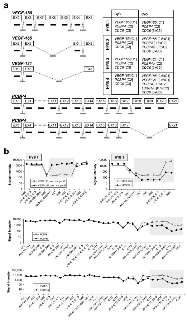
Figure 2.
VEGF and PCBP4 isoforms in the pilot experiment. (a) Simplified organization of VEGF and PCBP4, showing the included exon (black) and junction (gray) probes, and the hybridization scheme used to evaluate the artificial splicing forms. (b) Detection of VEGF and PCBP4 isoforms in the pilot experiment. Probes with differences in signal intensities are indicated by shadowed boxes.

