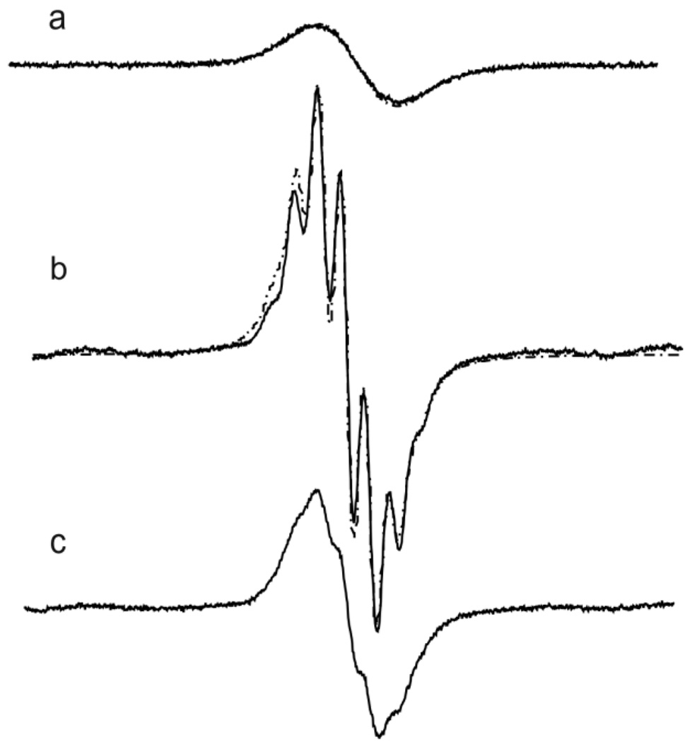
Figure 1.
X-band EPR spectra of 1 mM aTAMe (a), aTAMe* (b) and aTAMe** (c) measured in anoxic DMSO solutions radicals at room temperature. The spectra are shown with the same receiver gain. Spectral parameters were as follows: microwave power, 0.63 mW; modulation amplitude, 0.01 G; sweep width, 2G; number of points, 1024. The simulated EPR spectra (a) and (b) are shown by dotted lines.

