
Fig. 3.
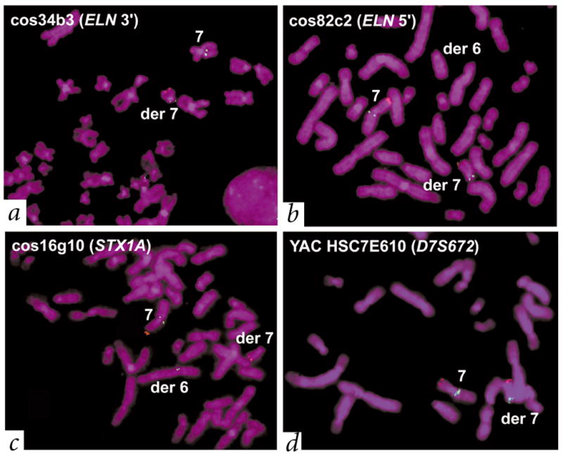
Individual with atypical WBS (11719) with a t(6;7)(q27;q11.23) translocation also carried the WBS inversion. The father (11976) of this affected individual also carries the inversion but not the translocation (Table 1). We used a control probe on chromosome 7p22 (RP11-13N3 containing LFNG, red) to identify the derivative chromosome 7. We used multiple test probes (green) to determine the site of the translocation and the extent of the inversion in patient 11719. a, b, We mapped the translocation breakpoint to the immediate 5′ end of ELN, on the basis of either the presence or absence of signals on the derivative chromosomes (see Fig. 1 for location; note that no deletions in the region were detectable). Characterization of the WBS inversion. c, d, We observed that probes cos34b3 and cos82c2 had the same pattern of hybridization to the translocation chromosomes as HSC7E610 (hybridizing to the derivative chromosome 7), whereas cos16g10 hybridized to the derivative chromosome 6. This suggests a probe order of HSC7E610 (D7S672)–cos34b3 (3′ ELN)–cos82c2 (5′ ELN)–cos16g10 (STX1A). On a normal chromosome, however, the known order of probes is 7cen–HSC7E610 (D7S672)–duplicon–cos16g10 (STX1A)–cos82c2 (5′ ELN)–cos34b3 (3′ ELN)–duplicon–7qter (see Fig. 1). Thus, patient 11719 carried the WBS inversion. The combined results of testing 20 probes using the same strategy indicate that the inversion breakpoints in this patient occurred within the duplicons.