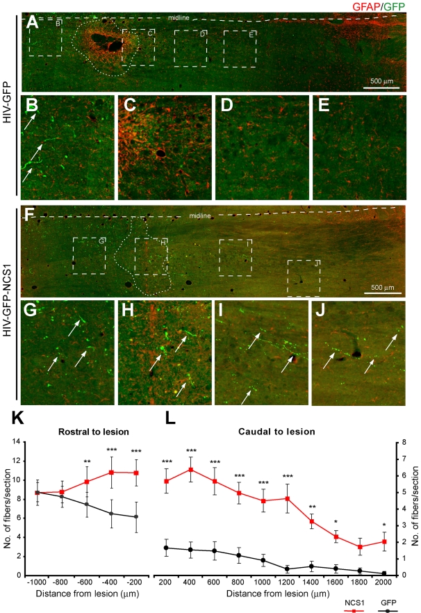
Figure 11. Delayed NCS1 overexpression after pyramidotomy induces axonal sprouting and regeneration from the lesioned CST.
Photomicrographs of GFP positive fibers in horizontal sections of the caudal medulla oblongata following a unilateral intracortical injection of lentivector 2 d after a pyramidotomy. Images are orientated in the rostral-caudal orientation from left to right. GFP positive fibers are depicted as green and the reactive astrocytes demarcating the lesion site are depicted as red. (A–E) In control HIV-GFP-transduced rats, GFP positive fibers (arrows) are seen approaching the rostral edge of the lesion (B) but few GFP positive fibers distal to the lesion. (F–J) In HIV-GFP-NCS1-transduced rats, GFP positive fibers (arrows) are seen approaching the lesion site from the rostral side. Equally important, GFP positive fibers (arrows) are present caudally with a distance of up to 2 mm distal to the lesion. (K–L) Quantification of GFP labelled fibers in the side ipsilateral to the pyramidal tract lesion demarcated by the midline with the lesion site as 0 µm. The HIV-GFP-NCS1-transduced rats showed a significant increase in the number of GFP positive fibers along the rostral and caudal regions to the lesion when compared with the control HIV-GFP-transduced rats. Panels B–E and G–J are higher magnifications of areas indicated by dashed boxes of panels A and F, respectively. Data are expressed as mean ± SEM from n = 4–5 rats per group with 5–6 sections per animal. * p<0.05, ** p<0.01, *** p<0.001, two-way ANOVA, Tukey post hoc test. Scale bar: 500 µm.

