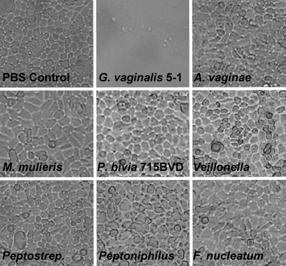
Fig. 3.
Cytotoxic changes of vaginal epithelial cell monolayers challenged with G. vaginalis strains and various BV-associated anaerobes. Bacteria were grown anaerobically in sBHIG at 37 °C for 24 h, and cultures were standardized to ensure equal numbers. Bacteria were added to vaginal epithelial cells, and incubated for 4 h. Light microscopy images were taken after incubation for 4 h. P. bivia strain BVD (not shown) did not exhibit cytotoxicity, and produced results that were similar to those of P. bivia strain 29303.

