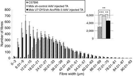
Figure 5.
Fiber diameter and fiber type distributions in U7-DYS/ sh-AcvRIIb-3 AAV-1-injected TA muscles. The tibialis anterior muscles of mdx mice were injected with 2 × 10¹¹ viral genome of either U7-DYS/sh-AcvRIIb-3 AAV or sh-control AAV. Muscles injected with AAV coding for sh-control are in gray and U7-DYS/sh-AcvRIIb-3-injected TA are in black. Three months after injection, mice (n = 9) were killed, and muscles were sectioned and labeled with laminin. The number and size of each fiber was measured. As an indicator, the data obtained with age-matched C57/Bl6 were added (hatched bars). **P < 0.01; ***P < 0.001. PBS, phosphate-buffered saline; TA, tibialis anterior.

