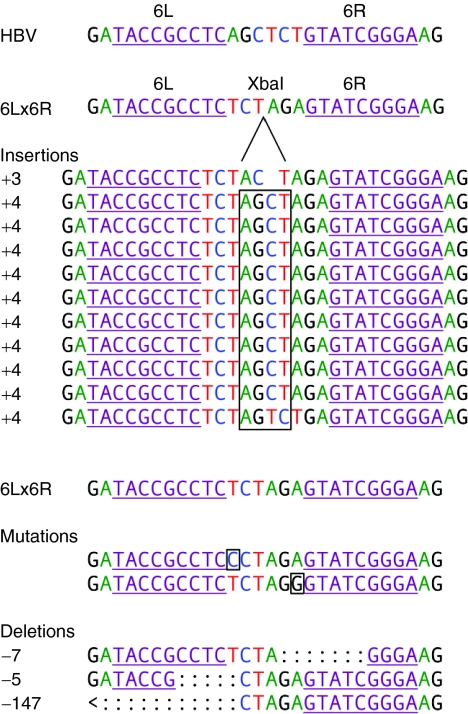
Figure 6.
Sequence analysis of imperfectly repaired ZFN-cleaved HBV DNAs. DNAs from cells co-transfected with 6L and 6R and the 6L/6R binding site were PCR-amplified with primers flanking the ZFN-binding site and digested with XbaI (Figure 5a, lanes 5–8). The reactions were further amplified with nested primers and cloned for sequencing in order to determine the types of mutations present. The ZFN 6L and 6R binding sites are underlined in each sequence and in the target site in HBV and pTHBV2 (first line) and in the XbaI-containing 6L/6R target (second line). The insertions are listed next to the predominant 4 base-pair (bp) insertion, and the frameshift is boxed. A second 6L/6R target sequence is listed above the mutations (middle) and deletions (bottom). The 4 bp insertions and first two deletions would result in frameshifts and large truncations in the core protein–coding region of the genomic HBV DNA. The last large deletion would truncate the upstream precore protein and delete the start codon for the core protein. HBV, hepatitis B virus; ZFN, zinc-finger nuclease.

