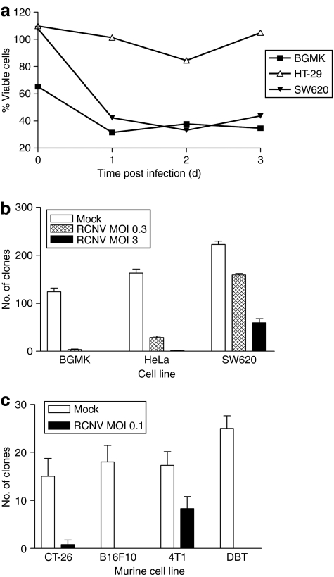
Figure 2.
Tumor cell killing by RCNV. (a) Cell killing in response to virus infection. The indicated cell lines were seeded in 6-well plates and RCNV added at an MOI of 3. At each of the indicated days after infection, the cells were collected and viability determined by Trypan blue exclusion using a Vi-CELL machine. This viability was then normalized to uninfected control cells. (b) Clonogenic assays to determine cell death induced by RCNV. The indicated cell lines were infected at an MOI of 0.3 or 3, and 24 hours after infection, trypsinized and counted. 103 viable cells were added in quadruplicate to 12-well plates and allowed to grow into visible colonies, which were visualized using 0.1% crystal violet. (c) Murine cells were infected with RCN-gfp at an MOI of 0.1 and clonogenic assay performed as above 3 days after infection. MOI, multiplicity of infection; RCNV, Raccoonpox virus.

