Figure 3.
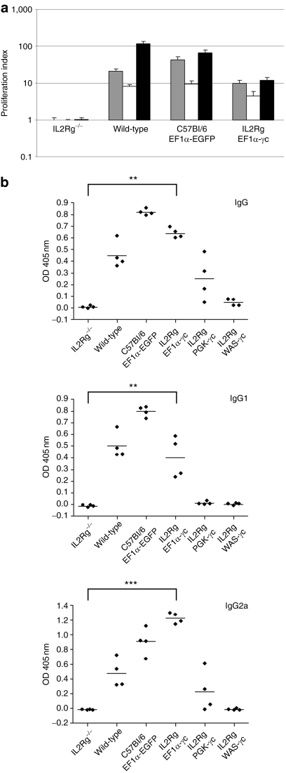
Restoration of immune function following lentiviral vector–mediated gene transfer. (a) Splenocytes from transplant recipients or wild-type mice were stimulated under conditions to promote T-cell proliferation. Proliferating cells were evaluated by the incorporation of [3H] thymidine and expressed as the ratio of counts obtained for stimulated to unstimulated cells. Gray bars, conA alone; white bars, IL-2 alone; black bars, conA and IL-2. (b) Humoral immune responses, indicated by serum IgG, IgG1, and IgG2a levels, were examined in mice transplanted with vector-treated C57Bl/6 or IL2RG−/− progenitors, and compared to γc−/−Rag2−/−c5−/− mice transplanted with untransduced IL2RG−/− cells. Histograms represent the mean value for each group (n = 4) with error bars representing the standard error of the mean. *P < 0.05, **P < 0.01, ***P < 0.0001 (Wilcoxon rank-sum test). EF1α, elongation factor-1-α OD, optical density; PGK, phosphoglycerate kinase; WAS, Wiskott–Aldrich syndrome.

