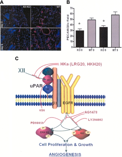
Figure 7.
Angiogenesis in FXII KO mice. (A left) Representative immunofluorescent stain with anti-CD31 of the punch biopsies at 20× magnification of skin at days 0 and 9 of the FXII KO or wild-type mice. (B) Bar graph of the means ± SDs of 4 or more slides of anti-CD31–stained vessels (platelet endothelial cell adhesion molecule) in the FXII KO or wild-type (WT) mice at day 0 (0) or day 9 (9). (C) Model of FXII signaling in HUVECs. FXII binds to domain 2 of uPAR on HUVEC membranes. Binding is blocked by plasma HKa or peptide HKH20 from domain 5 of HK or LRG20 from domain 2 of uPAR. FXII engagement induces uPAR's communication to the cell through a β1 integrin. Mab 6S6 to β1 integrin blocks intracellular signaling. Cell stimulation through the integrin or uPAR is mediated through the EGFR because the specific EGFR inhibitor AG1478 blocks FXII-initiated signaling. The MEK inhibitor PD98059 blocks FXII-induced ERK1/2 phosphorylation. Alternatively, LY294002, a PI3 kinase inhibitor, blocks FXII-induced Akt phosphorylation. Cross talk between ERK1/2 and Akt systems also occurs. Inhibition of either of these pathways blocks cell growth, proliferation, or angiogenesis arising from FXII treatment of HUVECs and aortic segments.

