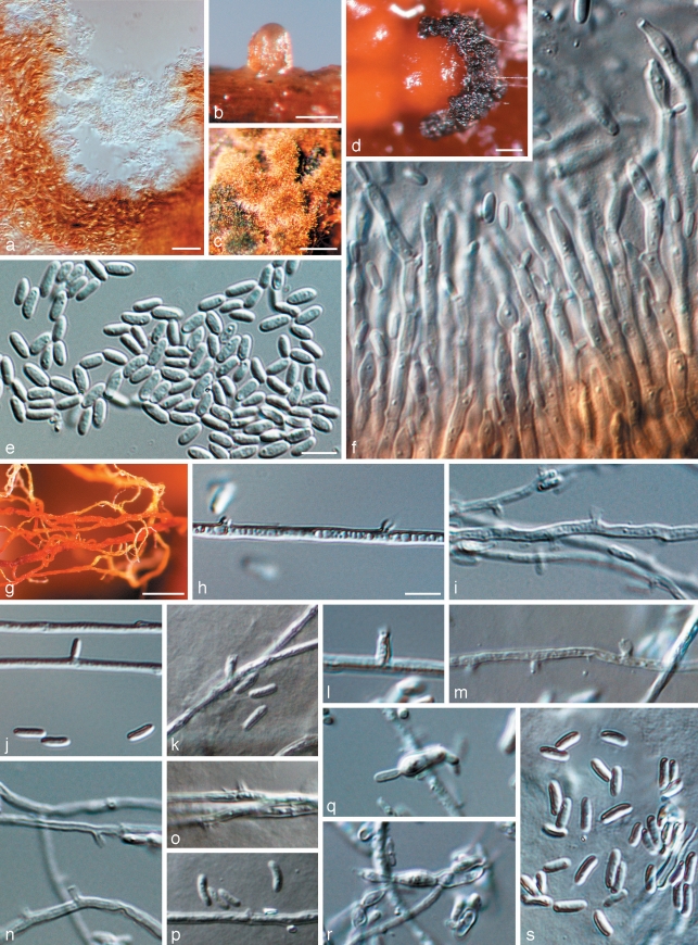
Fig. 6.
Collophora rubra. a. Longitudinal section through a pseudopycnidium; b–d. pseudopycnidia on pine needle (b), grapevine wood (c) and MEA medium (d); e. conidia formed in pseudopycnidia; f. conidiophores lining the inner wall of a pseudopycnidium; g. hyphae on PDA medium; h–p. conidiogenous cells on hyphal cells; q–r. microcyclic conidiation; s. conidia formed on hyphal cells. All from ex-type culture CBS 120873. a, e, f, h–s: DIC; b–d, g: DM. — Scale bars: a = 20 μm; b, c = 100 μm; d, g = 200 μm; e, h = 5 μm; e applies to e, f; h applies to h–s.

