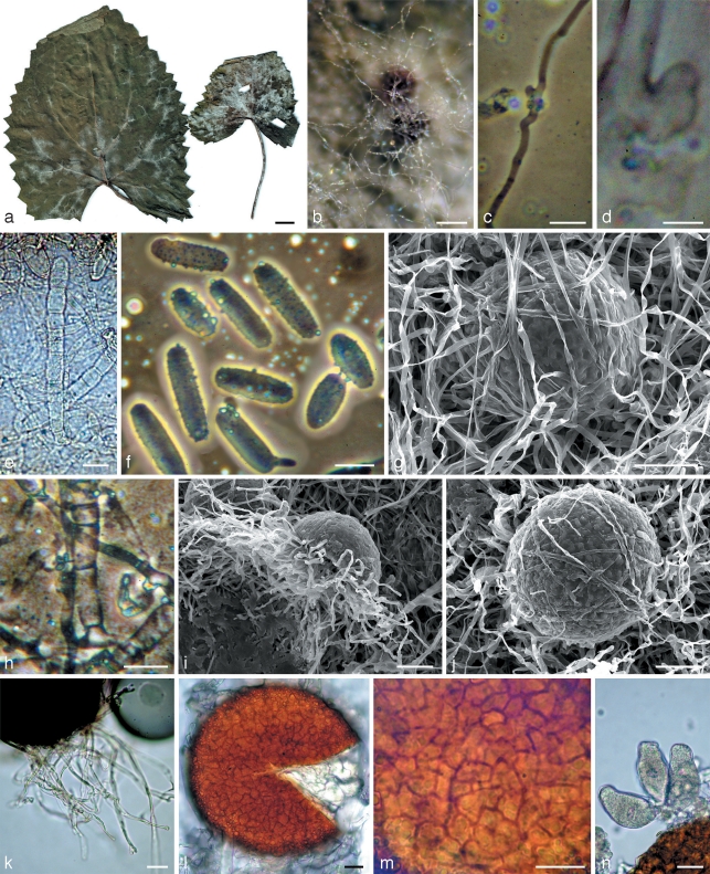
Fig. 3.
Morphology within phylogenetic subclade B4. a, b, g, i–n: Neoerysiphe hiratae (isotype, KW 34787F) on Ligularia stenocephala; c–f, h: N. hiratae (KW 34783F) on L. delphiniifolia. a. The infected host; b. chasmothecia in reflected light covered by the secondary mycelium; c, d. hyphae of the primary mycelium with appressoria; e. conidiophores; f. conidia; g, i, j. chasmothecia viewed by scanning electron microscope: g, j – covered by hyphae of the secondary mycelium, i – side view; h. basal part of conidiophore; k. chasmothecial appendages; l. chasmothecium viewed by light microscope; m. peridial cells; n. asci. — Scale bars: a = 1 cm; b = 100 μm; c, e, f, h, k–n = 20 μm; d = 5 μm; g, i, j = 50 μm.

