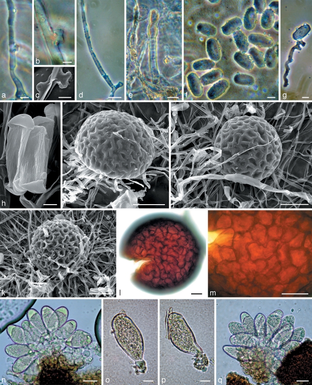
Fig. 5.
Morphology within phylogenetic subclade C4. a, b, d–g, q: Neoerysiphe nevoi (KW 35726F) on Thrincia tuberosa; c, h–j, l–p: N. nevoi (holotype, KW 34802F) on Tolpis virgata; k: N. nevoi var. scolymi (holotype, KW 34800F) on Scolymus hispanicus. a–c. Hyphae of the primary mycelium with appressoria; d. secondary hypha arisen from the primary hypha; e. conidiophore; f–h. conidia; g. germinated conidium with a hypha extending from a lobed appressorium of the Striatoidium type; i–k. chasmothecia viewed by scanning electron microscope; l. chasmothecium viewed by light microscope; m. peridial cells; n–q. asci. — Scale bars: a, f, g, o, p = 10 μm; b, c, h = 5 μm; d, e, l–n, q = 20 μm; i–k = 50 μm.

