Figure 1.
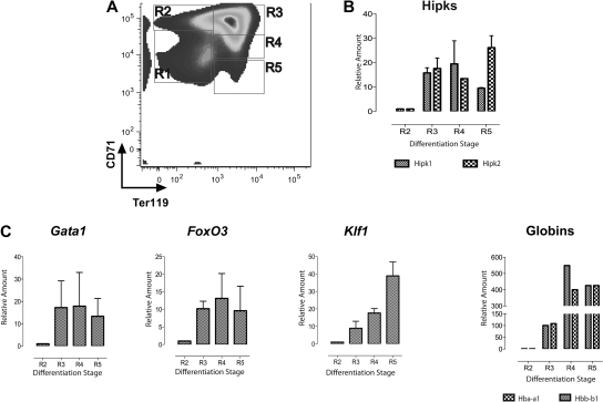
Induction of Hipk1 and Hipk2 during in vivo erythropoiesis. (A) Freshly isolated E14.5 murine fetal liver cells analyzed by FACS with antibodies staining for CD71 and Ter119. Regions R1 through R5 are defined by their staining patterns: CD71medTer119low, CD71highTer119low, CD71highTer119high, CD71medTer119high, and CD71lowTer119high, respectively. (B) Quantitative RT-PCR on mRNA isolated from each corresponding stage in panel A using primers against Hipk1 and Hipk2. (C) Quantitative RT-PCR using primers against other erythroid-specific genes: GATA1, FoxO3, Klf1, and α (Hba-a1)– and β (Hbb-b1)–globins. The relative amounts shown for each transcript are compared with the level of the corresponding transcript in R2 cells; n = 3 (mean ± 2 SEM).

