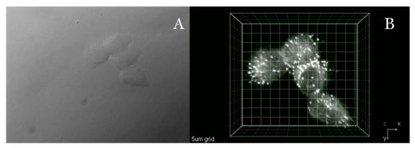
Figure 6.
Two-photon fluorescence micrograph of HCT 116 cells incubated with probe I (20 μM, 1 h 50 min). A) DIC, B) 3D reconstruction from overlaid 2PFM images obtained from a modified laser scanning confocal microscopy system equipped with a broadband, tunable Ti:sapphire laser (220 fs pulse width, 76 MHz repetition rate), pumped by a 10 W frequency doubled Nd:YAG laser. (60x objective, NA= 1.35). Scale: 5 μm grid.

