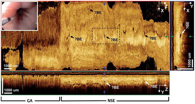
Fig. 6.

3D-OCT orthoplanes of distal esophagus previously treated with radiofrequency ablation (RFA). The en face XZ orthoplane of the epithelium at 390 μm tissue depth shows scattered, buried glands (?Barrett’s esophagus [?BE]) beneath neo-squamous epithelium. The cross-sectional YZ or-thoplane shows gastric (GA) and neosquamous epithelium (NSE) regions. The cross-sectional XY orthoplane shows scattered glands buried 350 –400 μm beneath the tissue surface. Inset Video endoscopy image.
