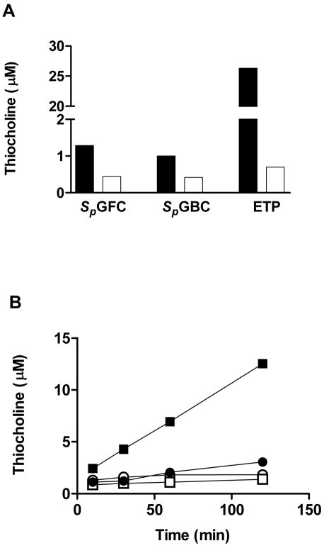
Figure 4.
(A) Thiocholine formation as detected by the BES-Thio method from the hydrolysis of different OPs (SpGFC, SpGBC and ETP) incubated with G117H (filled bars) or wild type BuChE (open bars) for 4 h. The OP concentration was 0.4 mM, and the enzyme concentration was 0.43 μM. (B) Thiocholine formation from oxime-mediated SpGBC hydrolysis by wild type AChE (HI-6 (■) and 2-PAM (●) and wild type BuChE (HI-6 (□) and 2-PAM (○)) as detected by the BES-Thio method. In the incubation system, AChE or BuChE was 0.25 μM, SpGBC was 0.1 mM, and oxime (2-PAM or HI-6) was 0.5 mM. The incubation times were 10, 30, 60 and 120 min, respectively.

