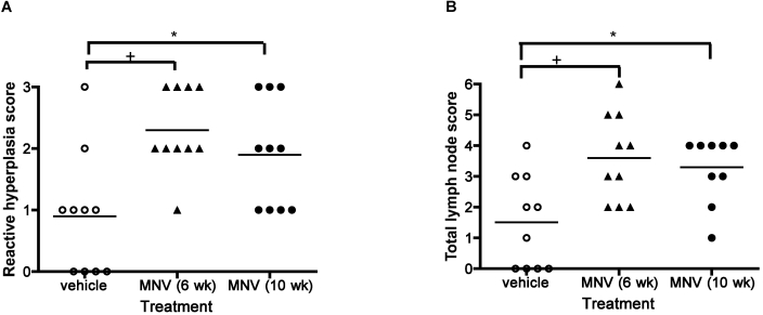
Figure 7.
Pathology of MLN in mice fed HFD. MLN of mice fed HFD for 10 wk were stained with hematoxylin and eosin and analyzed morphologically by a veterinary pathologist blinded to treatment group. (A) MNV infection was significantly (one-way ANOVA, P = 0.0034) associated with increased reactive hyperplasia. A multiple comparisons test found significant (*, P < 0.05; +, P < 0.01) differences between infected and uninfected groups. (B) The total lymph node score was significantly different between 3 groups (one-way ANOVA, P = 0.0033). Horizontal bar indicates the mean value. A posthoc test (Bonferroni) showed that the MNV-uninfected group was significantly different from the MNV-infected group (*, P < 0.05; +, P < 0.01)

