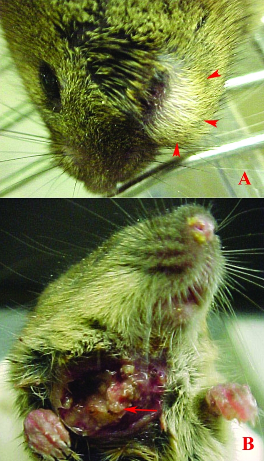
Figure 1.
(A) Clinical presentation of a typical maxillofacial abscess in a mouse, with swelling inferior to the eye (arrowheads). (B) Clinical presentation of a mandibulofacial abscess in a mouse, with drainage ventral to the mandibular ramus. Note the multifocal, well-defined necrotic nodules of the botryoid abscess (arrow).

