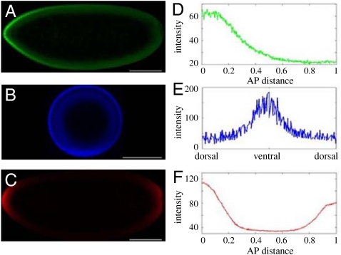
Fig. 1.
Maternal morphogen gradients in the early embryo. See ref. 7 for the method used to quantify the profiles. Embryos imaged for (A) Bicoid, (B) Dorsal, and (C) phosphorylated MAPK. (Scale bar: 100 μm.) (D) Gradient of Bicoid along the scaled AP axis. (E) DV gradient of nuclear dorsal. (F) Phosphorylation gradient of MAPK with peaks at the termini of the embryo.

