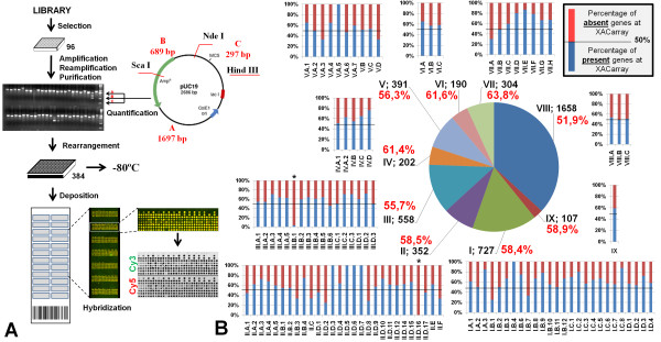
Figure 2.
(A) Figurative fluxogram of the XACarray construction protocol. The clones were selected from the library, amplified and re-amplified. The PCR products were purified and visually analyzed for concentration based on a molecular weight standard created specifically for this purpose and by re-sequencing of one of the ends of the insert. The samples were rearranged in 384 well plates and fixed onto the slides. The image demonstrates the result obtained with the hybridization of the DNA samples labeled with the fluorophores Cy3 and Cy5, highlighting subarray 7, for which the monochromatic analysis data demonstrates the quality of the hybridization. (B) Statistical distribution of the products fixed on the XACarray based on primary, secondary and tertiary annotation categories [6]. Note that for only two subcategories (III.B.1 and II.D.16), no products were represented on the XACarray, and in most of the remaining categories, the total of the genes present (blue bars) exceeds the absent genes (red bar).

