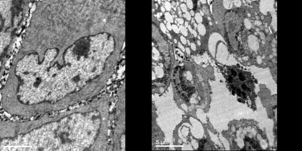
Figure 4.

The morphologic changes of each group cells observed by electromicroscope. Antisense group showed more cell degeneration and necrosis, with cell volume enlargement, chromatin margination and dissolving and lipid droplets within the cytoplasm increase, endoplasmic reticulum dilation, and swelling of mitochondria like vacuoles. Occasional plasma membrane rupture and cell collapse were also seen. (a: control group(original magnification × 10000 b: antisense group(original magnification × 4000).
