
Figure 2. Upregulation of c-mip in lymph nodes of eight patients with cHL-MCNS is restricted to Hodgkin and Reed-Sternberg (HRS) cells.


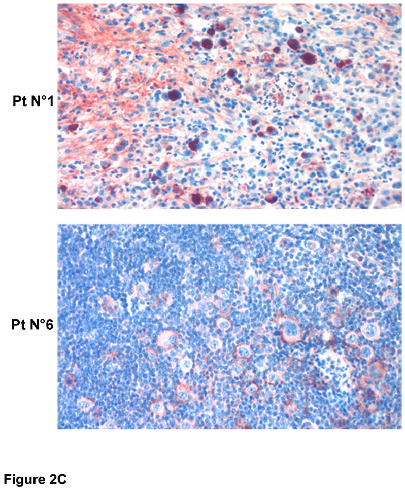

A, Immunohistochemistry analysis of c-mip in normal lymphoid tissues (thymus, lymphoid follicle of the spleen and lymph nodes); note that c-mip is not seen in normal tissues, except in the transitional zone of node tissue and in the lymphoid follicle (indicated by arrows) (original magnification, X20); B, Representative image showing c-mip in lymphomatous tissues from two patients with isolated cHL (original magnification, X20) (HRS cells are indicated by arrows); note that c-mip is undetectable in lymphomatous tissues from isolated cHL; C, Representative expression of c-mip in lymphomatous tissues from two patients with cHL-MCNS (original magnification, X20). c-mip shows intense staining in cHL-MCNS, restricted to HRS cells. D, Localization of c-mip in HRS cells from four patients with cHL-MCNS; no staining was detected in the HRS cells of two patients with isolated cHL (original magnification, X100). C-mip upregulation in HRS cells was found in all patients with cHL-MCNS, whereas no immunostaining was observed for c-mip in 9 control cases with isolated cHL