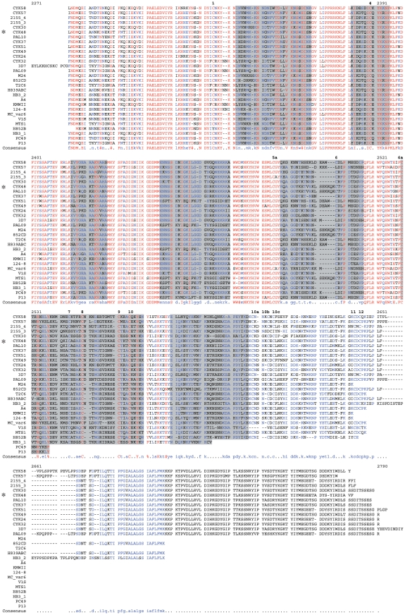
Figure 1. Alignment of the entire sequence of the DBL6ε domain from various isolates.
With respect to the 3D7 PFL0030c residue numbering and combined the alignment, ID5 is located from M2287 to K2328, DBL6ε from G2329 to P2651, transmembrane domain from N2677 to P2710 and ATS from K2711 to the C-terminus. The highly conserved amino acids are coloured in red, the lowly conserved amino acids are in blue, and the highly variable amino acids are in black. The seven polymorphic blocks are highlighted in gray. At the top of the sequences, cysteine residues are numbered from 1 to 12 in bold. (*) indicates the sequence of the expressed and purified CYK48-DBL6ε domain. On the left, all strains and isolates are presented; all are listed in Table 1.

