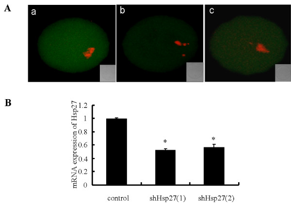
Figure 1.
Ad-shHsp27 effects specific suppression of Hsp27 expression in mouse oocyte. (A) Immunofluorescence detection of Hsp27 expression after Ad-shHsp27 infection for 48 h. Oocytes were fixed in 4% paraformaldehyde and then stained with anti-Hsp27 antibody (green). Chromosome material was counterstained with propidium iodide (red). a) control-infected oocyte. b) shHsp27(1)-infected oocyte. c shHsp27(2)-infected oocyte. (B) The results of real time RT-PCR showing Ad-shHsp27 infection for 24 h in mouse oocytes. The expression level was calculated from the Ct values by the 2(-Delta Delta Ct) method, and the mRNA ratio (arbitrary units) of Hsp27 was calculated with respect to that of control. Bar graphs indicate mean ± SD of four replicates. *P < 0.05 vs. control.

