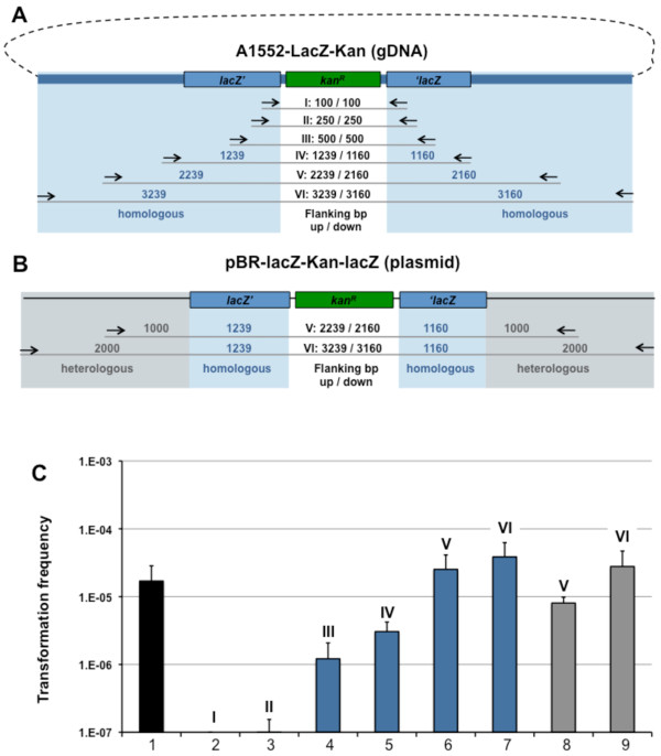
Figure 3.
PCR-derived donor DNA with various lengths of homologous and heterologous flanking regions. Panel A: PCR-derived fragments using genomic DNA of strain A1552-LacZ-Kan as template. Priming region of oligonucleotides (Table 1) are indicated. Primer combinations used: (I) KanR-100-flank-up & KanR-100-flank-down; (II) KanR-250-flank-up & KanR-250-flank-down; (III) KanR-500-flank-up & KanR-500-flank-down; (IV) NheI-lacZ-start & LacZ-end-SalI; (V) Tfm-II-gDNA-1000 & Tfm-II-gDNA+1000; (VI) Tfm-II-gDNA-2000 & Tfm-II-gDNA+2000. Panel B: Plasmid pBR-lacZ-Kan-lacZ was used as PCR template and is shown in a linearized fashion. The following primer pairs were used: (V) Tfm-II-1000 & Tfm-II+1000; (VI) Tfm-II-2000 & Tfm-II+2000. Sizes of the up- and downstream flanking regions with respect to the Kanamycin resistance cassette are indicated in the middle. Blue shading: region homologous to recipient strain (A1552; wild-type); grey shading: heterologous region. Panel C: V. cholerae wild-type strain A1552 was naturally transformed using the crab-shell transformation protocol and PCR-derived DNA according to Panels A and B. Transformation frequencies are shown on the Y-axis using either 2 ug gDNA of strain A1552-LacZ-Kan as positive control (lane 1; black) or 200 ng of PCR-derived DNA with varying length of the homologous (in blue; lane 2 to 7; according to Panel A) or homologous + heterologous (in grey; lane 8 and 9; according to Panel B) flanking region. Length of KanR-flanking DNA: lane 2: 100 bp, lane 3: 250 bp; lane 4: 500 bp; lanes 5: ~1000 bp; lane 6 and 8: ~2000 bp; lane 7 and 9: ~3000 bp. Average of at least three independent experiments.

