Figure 1.
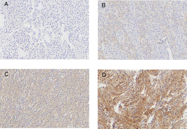
Immunohistochemical staining intensity scores. A) Meningiomas stained with anti-EGFR antisera showing negative staining. B) 1+ staining C) 2+ staining D) 3+ staining. Original magnification for all images was 40 ×.
Images are arranged as follows: Upper left (A), upper right (B), lower left (C), lower right (D).

