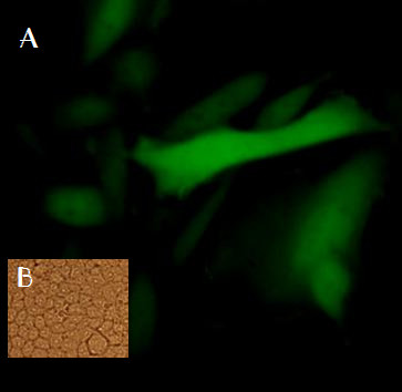
Figure 3.

A. CHO cells transfected by EGFP-λ-phage nanobioparticles using fluorescent microscopy. Four cell types COS-7, CHO, TC-1 and HEK-293 were transfected by EGFP-λ-phages. The best GFP expression was observed after 48 hours by fluorescent microscopy 48 hours later in CHO cells. B. CHO experimental control.
