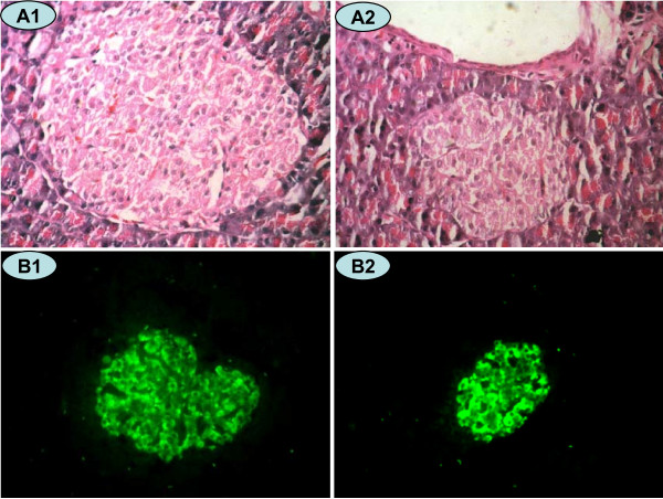
Figure 3.
HE Staining (×400 magnification) and insulin immunofluorescence staining (×200 magnification) in a rat pancreatic islet. Islet cells in the NC (A1 and B1) and CUGFR (A2 and B2) groups 4 weeks after catch-up growth with HE staining (A1 and A2) and insulin fluorescence staining (B1 and B2). Green fluorescence shows cells positive for insulin.

