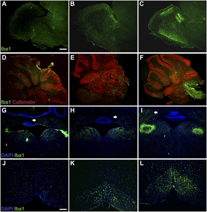
Fig. 2.
Progressive inflammatory response in the brains of KO mice. (A–C) Microglial activation in the OB of KO mice at early (A), middle (B), or late (C) stage of the disease was determined using an antibody against Iba-1.(D-F) Calbindin and Iba-1 staining in the cerebellum of KO mice at early (D), middle (E), or late (F) stage of the disease. (G–I) Iba-1 staining in the vestibular and deep cerebellar nuclei of KO mice at early (G), middle (H), or late stage (I) of the disease. (J–L) Iba-1 staining in the IO of KO mice at early (J), middle (K), or late stage (L) of the disease. Iba-1 staining in the brains of KO mice at different stages of the disease (early, middle, and late stages; n = 5 each) shows a localized and progressive inflammatory response. In early-stage animals (A, D, G, and J), marked microglial activation is already present in the OB (A), VN (G), and deep cerebellar nuclei (G, arrow). However, no microglial response is visible in either the cerebellum (D) or in the vicinity of the IO (J). In middle-stage animals (B, E, H, and K), enhanced microglial accumulation is observed in the OB (B), VN (H), and, to a lesser extent, in the deep cerebellar nuclei (H, arrow), cerebellar lobes (E), and IO (K). In late-stage animals (C, F, I, and L), severe microglial activation is observed in the olfactory lobe (C) and the deep cerebellar nuclei (I, arrow). Extensive microglial reactivity along with localized tissue destruction is observed in the cerebellar lobes (F, where loss of calbindin staining is visible in the affected lobes) and VN (I). In contrast, only moderate activation is present surrounding the IO (L). (Scale bar, A–I.125 μm; J–L, 50 μm.)

