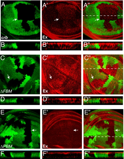
Fig. 6.
Loss of crb results in mislocalization of Ex in imaginal disk epithelial cells. In all panels, wing discs were stained with α-Ex antibody (red) and mutant clones (GFP-negative) are indicated with arrows. (A–A″) A horizontal section through a wing disk containing crb clones. The optical section was captured midway down from the apical surface of epithelium. Note increased Ex staining in crb clones. The dotted line in A″ indicates the position of vertical section in B–B″. (B–B″) A vertical section through the wing disk in A–A″ (apical is to the top). Ex localization extended more basolaterally in crb clones. (C–D″) Similar to A–B″ except that crbΔFBM clones were analyzed. Note the mislocalization of Ex to more basolateral position in crbΔFBM clones. (E–F″) Similar to A–B″ except that crbΔPBM clones were analyzed. Note the relatively normal apical localization of Ex in crbΔPBM clones.

