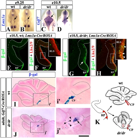
Fig. 2.
A switch in cell fate causes RP reduction in dreher mice. (A–D) Wild-type (A and C) and dreher (dr/dr; B and D) embryos stained with RP markers. Arrows point to fourth ventricle RP, which is induced in dreher embryos but does not grow properly. (E–H) Lineage analysis of RP cells in wild-type (E and F) and dreher (G and H) embryos using the Lmx1a-Cre/ROSA alleles. Insets (F, H) show higher magnification of RLS. In dreher mutants, some RP cells lose their identity and migrate into the cerebellar anlage (G, arrow). These β-gal+ cells could be labeled by an anti-Lhx2/9 antibody (Inset in H, arrowheads), which specifically marks RLS cells (5). (I and J) Lineage analysis of RP cells using the Gdf7-Cre/ROSA alleles. Sagittal sections of adult wild-type (I) and dreher (J) vermis stained for β-gal activity. In the wild-type Gdf7-Cre/ROSA cerebellum, β-gal staining is limited mostly to the CP (i′). In the dreher;Gdf7-Cre/ROSA cerebellum, ectopic β-gal+ cells are present in DCN (arrowheads) and IGL of the posterior vermis (arrow) (j′). (K) Diagram summarizing a switch in fate of RP cells in dreher mice. In wild-type mice, RP (red) produces CP. In dreher mice RP aberrantly produces neurons of DCN and granule cells and UBC of posterior vermis. (Scale bar: A and B, 800 μm; C and D, 1.75 mm; E–H, 180 μm; I and J, 870 μm; i′ and j′, 410 μm.)

