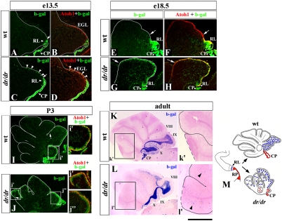
Fig. 4.
Abnormal distribution of granule cells originating from Lmx1a+ progenitors in the dreher cerebellum. Midsagittal sections of wild-type (A, B, E, F, I, and K) and dreher (C, D, G, H, J, and L) cerebella containing the Lmx1a-Cre/ROSA fate-mapping system at the indicated stages. Sections were stained with indicated antibodies (A–J) or were stained for β-gal activity (K and L). (A–D) Arrows point to the anterior limit of the RL in wild-type (A) and dreher (C) embryos. Arrowheads point to numerous β-gal+ cells migrating along the dorsal surface of the dreher (C and D) but not wild-type (A abd B) cerebellar anlage at e13.5. (E–J) Arrows indicate the anterior limit of β-gal+ cells in wild-type and dreher EGL. At both e18.5 and P3, β-gal+ cells extend much more into the anterior cerebellum in dreher mice than in their wild-type littermates. At all developmental stages, β-gal+ cells in both wild-type and dreher EGL express Atoh1 (D, F, H, I, i′, J, j′, and j′′), indicating that they are granule progenitors. (K and L) In adult wild-type vermis, β-gal+ cells predominantly populate posterior lobes IX and X (K). In dreher mice, numerous β-gal+ cells abnormally populate the intermediate and anterior vermis (L). Arrowheads (l′) point to ectopic β-gal+ cells in anterior IGL in ′adult dreher mice. (M) Diagram summarizing distribution of granule cells originating from Lmx1a+ RL progenitors in wild-type and dreher mice. Normally these cells (blue circles) contribute to posterior vermis. In dreher mice they overmigrate into intermediate and anterior vermis. (RP derivatives are shown in red. Fig. 2K gives a detailed description). (Scale bar: A–D, 290 μm; E–H, 530 μm; I and J, 420 μm; i′ and j′, 200 μm; K and L, 1.5 mm; k′ and l′, 750 μm.)

