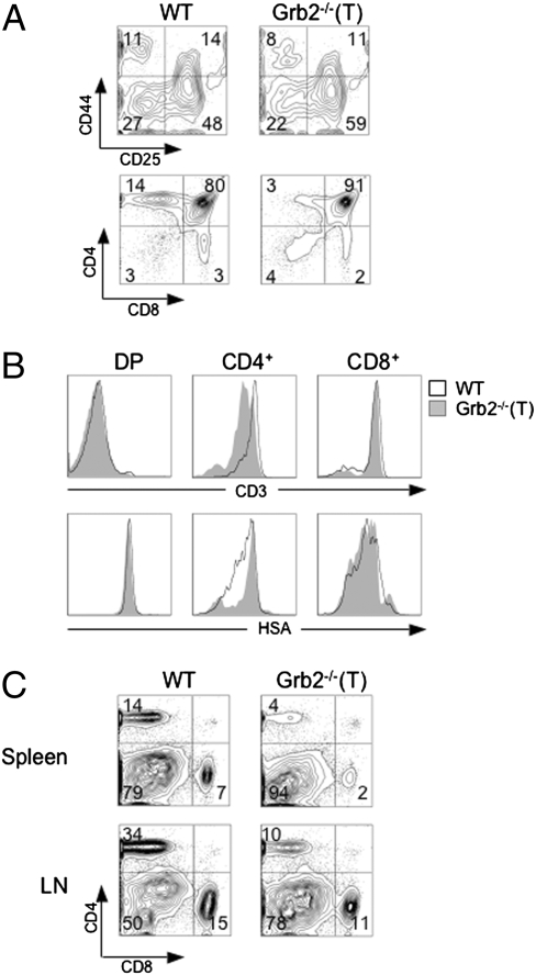
Fig. 1.
Impaired thymocyte development in Grb2−/−(T) mice. (A) Cellularity and CD4 and CD8 expression of Grb2−/−(T) thymocytes. Total thymocytes stained with anti-CD4 and anti-CD8 (Lower), or gated DN (CD4− CD8− B220− Gr1− CD11b−) thymocytes stained with anti-CD44 and anti-CD25 (Upper). Samples are from 8-week-old WT and Grb2−/−(T) mice. Total numbers of T cells and thymocyte subsets are given in Table S1. Results represent more than seven Grb2−/−(T) mice and WT mice (P < 0.01). (B) Cell surface expression of CD3 and HSA on thymocyte subsets. Shown are histograms of CD3 or HSA expression on gated DP, CD4+ and CD8+ SP cells. (C) Anti-CD4 and anti-CD8 staining of T cells from the spleen and lymph nodes (LN) of WT and Grb2−/−(T) mice. Results are representative of more than seven Grb2−/−(T) mice and WT controls (P < 0.01; Table S1).

