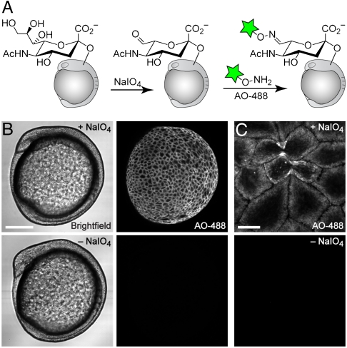
Fig. 3.
Imaging of sialylated glycans using a nonmetabolic approach. (A) Schematic for chemical labeling of sialylated glycans by treatment with NaIO4 to expose aldehydes on sialic acids, followed by detection using aminooxy-fluorophore conjugates. (B and C) Zebrafish embryos (10 hpf) were bathed in NaIO4 (500 μM, 30 min, top row) or no reagent (bottom row), reacted with aminooxy-Alexa Fluor 488 (AO-488, 100 μM, 1 h, pH 6.7), and imaged by confocal microscopy. (B, left panel) Brightfield; (B, right panel) and (C), maximum intensity z-projection images of AO-488 fluorescence. [Scale bars: 200 μm (B), 20 μm (C).]

