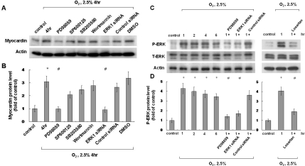
Figure 2. Effect of signalling pathway inhibitors on hypoxia-induced myocardin expression and ERK phosphorylation.
(A and B) ERK pathway mediates hypoxia-induced myocardin expression in neonatal cardiomyocytes. Neonatal cardiomyocytes were pre-treated with an ERK pathway inhibitor (PD98059), a JNK inhibitor (SP600125), a p38 MAPK inhibitor (SB203580), a PI3K/Akt inhibitor (wortmannin) or ERK siRNA, followed by hypoxia for 4 h. Neonatal cardiomyocytes were harvested and cell lysates were analysed by Western blotting using an anti-myocardin antibody. Result are normalized to actin levels. *P<0.01 compared with normoxia control; #P<0.01 compared with hypoxia for 4 h (n=3). (C and D) Hypoxia-induced phosphorylation of ERK in neonatal cardiomyocytes. Neonatal cardiomyocytes were subjected to normoxia or hypoxia for 1–6 h in the presence or absence of inhibitors, and cell lysates were collected for Western blot analysis using antibodies against total ERK (T-ERK) and phospho-ERK (P-ERK). *P<0.01 compared with normoxia control; #P<0.01 compared with hypoxia for 1 h (n=3).

