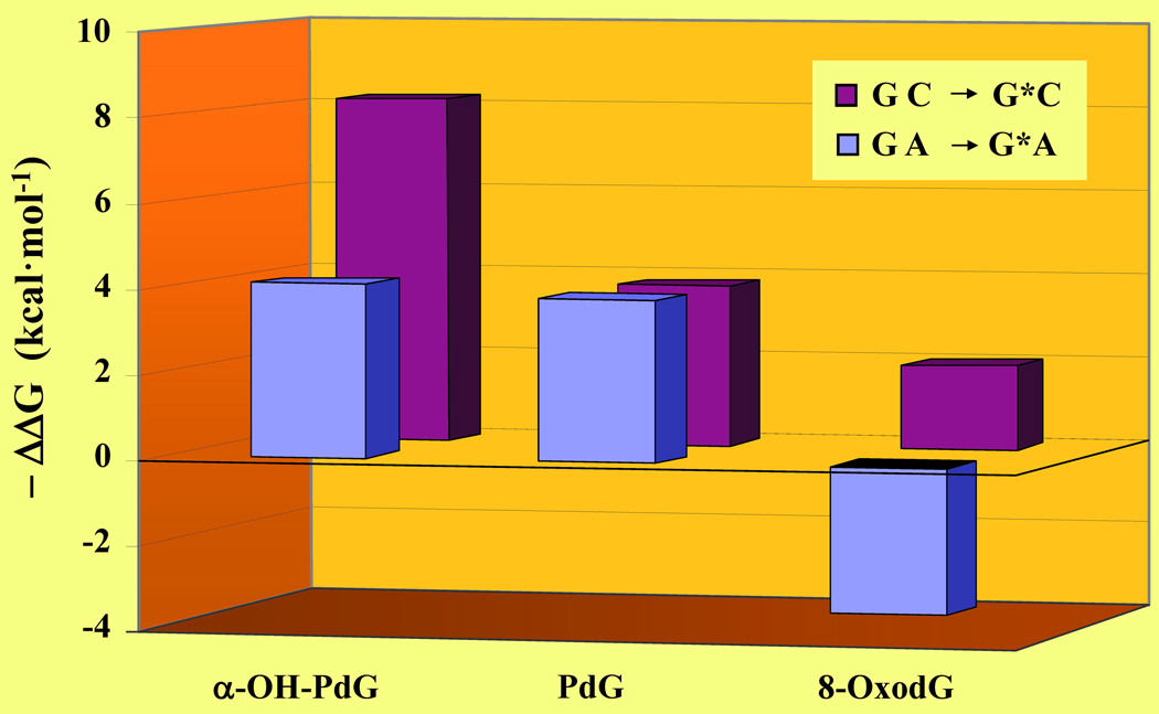
Figure 6. Lesion-Induced Energetic Impacts as a Probe of Cytotoxicity and Mutagenicity.
Comparison of the G to G* modification within the “nascent” and mismatch duplexes as monitored by the ΔΔG of G·C → G*·C (burgundy) versus G·A → G*·A (blue) for the α-OH-PdG, PdG,10 and 8-oxodG29 lesions. Although the thermodynamic parameters are derived from DNA duplexes of distinct sequence context under different solution conditions, each data set is obtained via direct comparison with its respective reference duplex (i.e., ΔΔGGC to G*C = ΔGG*C − ΔGGC and ΔΔGGA to G*A = ΔGG*A − ΔGGA). The lesion-induced impacts on duplex dissociation free energies are expressed as - ΔΔG to improve clarity and sorted on the basis of decreasing ΔΔG. Amongst these lesion-containing duplexes, 8-oxodG·A is thermodynamically stabilized relative to the corresponding “undamaged” G·A mismatch.

