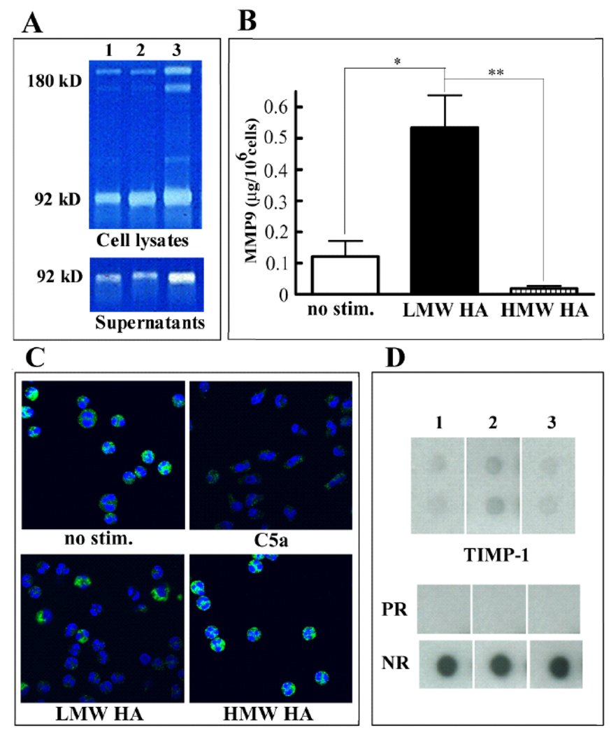
Figure 3.
Effect of HA on MMP-9 and TIMP-1 expression. A: Bone marrow cells were untreated (lane 1) or treated with 100 µg/ml HMW HA (lane 2) or LMW HA polymers (lane 3). Supernatants and lysates of the adherent layer were harvested and analyzed using zymogram gels. B: Neutrophils purified from the peripheral blood were unstimulated or exposed to 100µg/ml LMW HA or 100µg/ml HMW HA for 30 min. MMP-9 was detected in the cellular supernatants by ELISA (mean ±S.D, n=4–6). Differences between unstimulated cells and LMW HA-treated cells, and between HMW HA and LMW HA-treated cells were statistically significant (p=0.014 and p=0.001 respectively). C: Neutrophils were unstimulated or stimulated with 50nM C5a, 100µg/ml LMW HA or 100µg/ml HMW HA for 30 min as indicated and the intracellular expression of MMP-9 was determined by immunofluorescence. The nuclei were counterstained with DAPI. D: The expression of TIMP-1 in murine bone marrow cell supernatants was examined by using the RayBio Mouse Cytokine Antibody Array III&3.1. Where indicated, bone marrow cells were not treated (lane 1) or treated with HMW HA (lane 2) or LMW HA (lane 3). Dots indicated as PR show the positive reference and blank shows the negative reference (NR). The experiment was performed twice with similar results.

