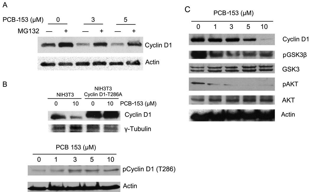
Fig. 4. Proteasome and T286 phosphorylation dependent pathways regulate cyclin D1 protein levels in PCB-153 treated cells.
(A) MCF-10A cells were treated with 50 µM of MG132 for 45 min followed by addition of PCB-153 for 4 h. Cyclin D1 protein levels were analyzed by immunoblotting. (B) Upper-panel: NIH3T3 mouse fibroblasts carrying wild type and T286A mutant form of cyclin D1 were incubated with 10 µM PCB-153 for 4 h. Cyclin D1 protein levels were analyzed by immunoblotting; γ-tubulin levels were used for comparison. Two-way ANOVA followed by paired t test were used to compare the two groups of treatment; n=3, p < 0.05. Bottom-panel: MCF-10A cells were treated with different doses of PCB-153 for 4 h and harvested for immunoblotting analysis of the T286 phosphorylated form of cyclin D1; actin levels were used for comparison. (C) Immunoblot analysis of cyclin D1, phosphorylated AKT and GSK-3β, total AKT and GSK-3β, and actin.

