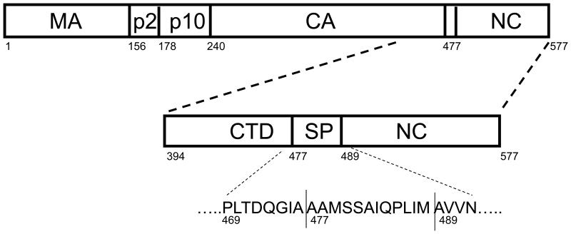
Figure 1.
Schematic representation of the RSV Gag polyprotein and the CTD-SP-NC polyprotein construct used in this study. CTD is from residue 394 to 474, SP from 475 to 488, and NC from 489 to 577. NC has two zinc fingers, ZF1 (509 – 522) and ZF2 (535 – 548) that together makeup the NC core (509 – 548). The horizontal lines in SP represent the PR cleavage sites between CTD and NC at 476 and 488. A third cleavage site also exists within SP at 479. The amino acid sequence comprising the “assembly domain” from residue 469 at the C-terminus of CTD to residue 492 at the N-terminus of NC is shown.

