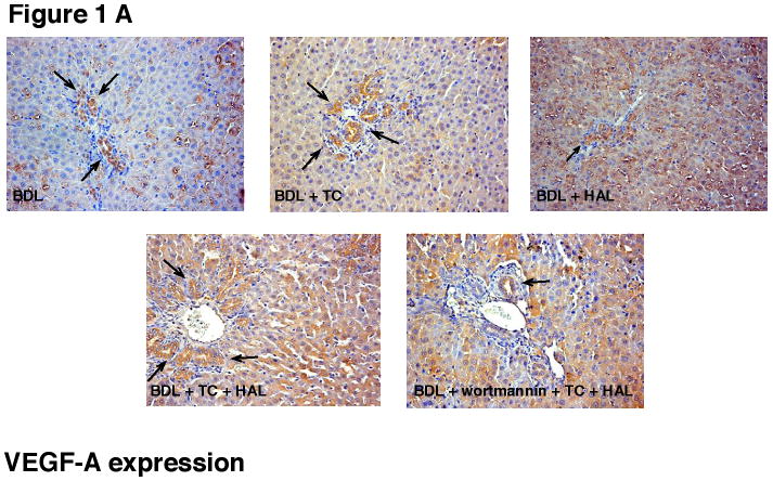
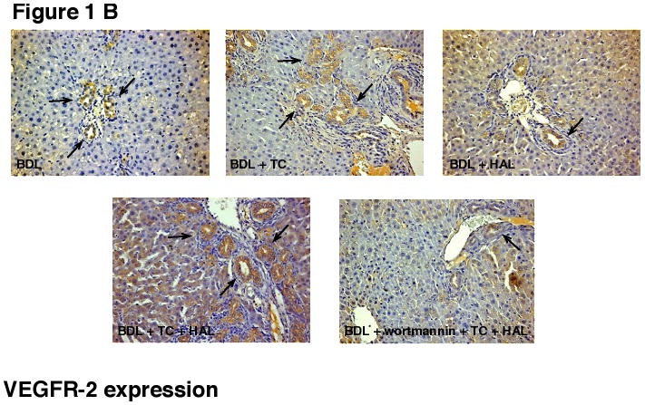
Figure 1.


Immunohistochemistry for [A] VEGF-A and [B] VEGFR-2 in liver sections from the selected groups of animals of Table 1. [A-B] Immunohistochemistry shows that bile ducts express VEGF-A and VEGFR-2 (arrows). HAL decreased the % of cholangiocytes positive for VEGF-A and VEGFR-2 (see Table 2 for semiquantitative analysis). Taurocholic acid feeding to BDL + HAL rats prevented HAL-induced decrease in VEGF-A and VEGFR-2 protein expression (see Table 2 for semiquantitative analysis), decrease that was blocked by the simultaneous administration of wortmannin (see Table 2). Original magnification 20×. VEGF-A = vascular endothelial growth factor-A; VEGF-R2 = vascular endothelial growth factor-receptor 2.
