
Fig. 6.
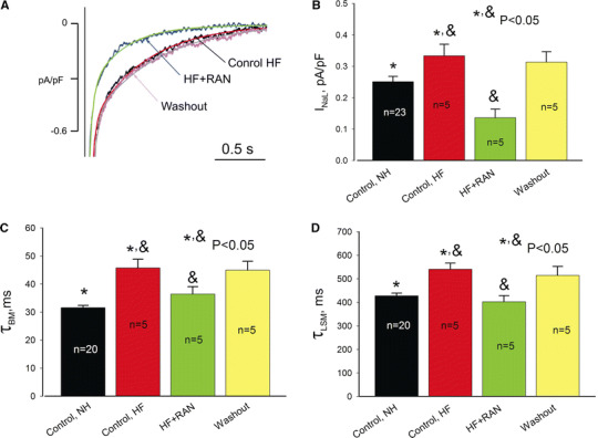
Effects of RAN (10 μM) on INaL in ventricular myocytes from canine failing hearts. a Representative traces along with double-exponential fit of the time course of INaL decay (solid lines). b Summary of the data on INaL density measured as a mean current within 200–220 ms after the depolarization onset, and decay kinetics (c, d) measured at −30 mV in control, in the presence of RAN, and during the washout. Data are mean ± SEM pooled from 5 to 23 cells. See details in text