
Fig. 1.
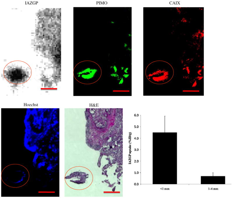
Comparison of intratumoral distribution of 131I-IAZGP with pimonidazole binding (green), hypoxia-regulated protein CAIX (red), and Hoechst 33342 (blue) in a tissue section containing disseminated intraperitoneal (i.p.) HT29 tumors. A larger tumor, part of which is seen in the upper right quadrant has low levels of 131I-IAZGP uptake, pimonidazole binding, and CAIX expression together with significant blood perfusion. The microscopic tumor with dimensions of ∼1 mm (circled) has high 131I-IAZGP uptake throughout the tumor together with high pimonidazole binding and CAIX expression. All images were obtained from the same tissue section. Scale bar =1 mm. The histogram shows quantitative 131I-IAZGP uptake in a collection of 27 i.p. HT29 tumors from a single animal expressed as % injected dose per gram. Tumor uptake was derived from an 131I-IAZGP autoradiograph with individual tumor sizes based on the H&E image. 131I-IAZGP uptake was significantly higher in microscopic i.p. tumors (<1 mm diameter, n=18) than larger ones (>1 mm, n=9), p<0.001