
Fig. 3.
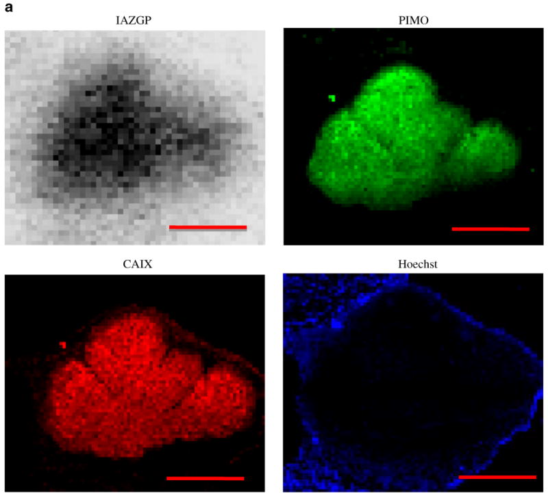
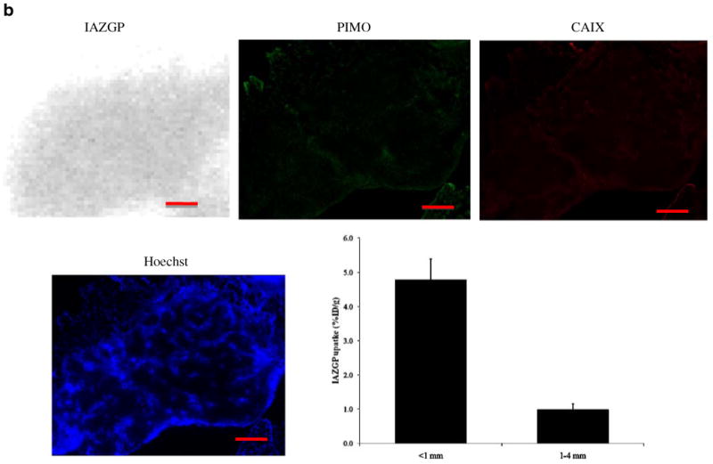
Comparison of intratumoral distribution of 131I-IAZGP uptake, pimonidazole binding, CAIX expression, and blood perfusion in a microscopic and a larger HT29 intradermal (i.d.) tumor. The microscopic tumor (a) shows high 131I-IAZGP uptake, pimonidazole binding, and CAIX expression with little to no blood perfusion. The larger tumor (b) shows the complete opposite in all respects. Scale bar=500μm. The histogram (b) shows a summary of quantitative 131I-IAZGP uptake in 11 HT29 i.d. tumors from a variety of animals. 131I-IAZGP uptake was significantly higher in microscopic i.d. tumors (<1 mm diameter, n=5) than larger ones (>1 mm, n=6), p<0.001